Dr. McDowell’s book is fascinating because he gives the history of our knowledge of trace minerals and how scientists and livestock producers described symptoms of both deficiencies and toxicities.
I think you might enjoy some of the history of trace mineral research.
Copper, molybdenum and sulfur
The necessity of copper for cattle was first established in the 1930s with the discovery in Florida that cattle that had a wasting disease were deficient in cobalt, iron and copper.
Researchers in northern Europe described this wasting disease by animals as having diarrhea, loss of appetite and anemia.
In the late 1930s, scientists in England described a severe scouring disease of cattle called “teart” that was caused by ingestion of forage with high levels of molybdenum.
Later scientists discovered that large doses of copper sulfate could prevent this condition. Still later, it was shown that molybdenum limited the retention of copper in the body, especially in the presence of adequate amounts of inorganic sulfate in the diet (water).
It was this discovery that led to numerous studies on the relationship among copper, molybdenum and sulfate. We can summarize these interrelationships as:
- Molybdenum in the presence of sulfate reduces the deposition of copper in organs and increases the excretion of copper in the urine.
- An increase of dietary copper reduces molybdenum deposition in the liver.
- When the copper-to-molybdenum ratio of forages in the presence of adequate sulfate was less than 2.8-to- 1, then copper deficiency is evident. A copper-to-molybdenum ratio of no less than 4-to-1 has been proposed to ensure the copper requirement will be met.
- High levels of dietary zinc and iron depress copper absorption and tend to increase the requirements. High dietary levels of zinc (100 ppm) reduce liver copper storage.
- Cattle can die from copper poisoning; these animals may experience nausea, vomiting, salivation, abdominal pain, convulsions, paralysis and death. The usual cause is improperly formulated supplements or diets.
Zinc
Zinc is widely distributed through the body, but animals have a limited ability to store zinc in a form that can be mobilized to prevent a deficiency.
In cattle, the highest concentrations of zinc were found in the following order: pancreas, liver, pituitary gland, kidney and adrenal gland. Additional reports have shown the testicles and accessory sex glands contain high concentrations.
It is suspected that zinc was applied for skin lesions by several cultures, including the Egyptians.
In 1960 scientists discovered that a skin disorder in cattle could be cured with zinc therapy.
Loss of appetite is one of the first signs of deficiency, and in calves, bowing of the hind legs and stiffness of the joints is noted.
In lab animals, severe zinc deficiency during pregnancy has resulted in offspring with impaired learning ability.
Additional clinical signs of a zinc deficiency in cattle include: inflammation of the nose and mouth with submucous hemorrhages, unthrifty appearance, rough hair coats, stiffness of the joints with swelling of the feet front of fetlocks, cracks in skin of coronary bands around the hooves and dry and scaly skin on the ears.
These findings contradict those stated in the petition to list the prairie dog as a threatened species. The impacts of these effects will depend on the goals of land managers.
MSU’s findings suggest that prairie dog colonies may not be ideal for grazing cattle due to a decrease in total standing crude protein.
Unlike other findings that suggest prairie dogs may have beneficial or neutral effects to native rangeland, the MSU study indicates that prairie dogs may have detrimental effects for some species.
In grazing animals, a marginal zinc deficiency results in subnormal growth, fertility, low serum zinc values, resistance to infection and stress.
Selenium
During the 1930s, selenium was identified as the toxic element in some forages that caused animals to lose hair, nails and hooves.
Selenium is known to be required by food animals and humans. Consumption of feedstuffs containing both toxic and deficient concentrations of selenium presents a problem for grazing livestock.
Marco Polo in his travels in western China (ca. 1295) described a syndrome resulting from the ingestion of seleniferous plants. He reported that when horses ate this poisonous plant, hooves dropped off.
In 1860 an army surgeon in South Dakota also described a fatal disease in horses grazing near Fort Randall. The horses exhibited extreme tenderness and inflammation of the feet, accompanied by loss of hair from the mane and tail.
Some speculate that many horses of the U.S. cavalry commanded by General Custer exhibited selenium toxicity during the summer of 1876.
Pioneers on the northern Great Plains in the 1890s also described selenium toxicity of their livestock. They associated the disease with alkali seeps and waters of high-salt content. It became known as alkali disease.
In the 1950s, selenium was reported to be beneficial for livestock and shown to prevent liver necrosis in swine and muscular dystrophy in calves.
Selenium is closely linked to vitamin E; both protect biological membranes from degeneration. Lack of these nutrients results in tissue breakdown.
Selenium deficiency in ruminants is called white muscle disease and is a degeneration of striated muscles. Animals with it have generalized weakness, stiffness, muscle deterioration and possible difficulty standing.
There are a couple of clinical patterns. The congenital type of muscular dystrophy in which calves are stillborn or die within a few days; or after sudden physical exertion such as nursing or running. It is observed in calves between 1 and 2 months old.
Poor reproductive performance also is a symptom of selenium deficiency and includes retained placenta. Work from Ohio showed that the incidence of retained placentas was reduced when cows were injected with a combination of selenium and vitamin E.
When consumed in sufficient amounts, alkali disease causes toxicity. Alkali disease generally happens when animals graze forages with selenium in the range of 5 to 40 parts per million (ppm). Certain selenium-mutating plants have between 100 and 9,000 ppm selenium.
Animals suffering from selenium toxicity have loss of appetite, lack of thriftiness, cirrhosis or the loss of hair, lameness and elongated hooves. ![]()
—Excerpts from extension.org
PHOTO
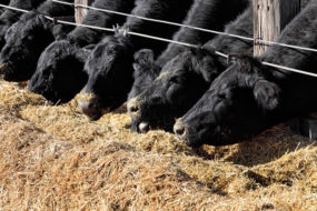
In grazing animals, a marginal zinc deficiency results in subnormal growth, fertility, low serum zinc values, resistance to infection and stress. Photo courtesy of New Generation Feeds.

John Paterson
Extension Beef Specialist
Montana State University
johnp@montana.edu


