It is a viral disease spread by biting midges, which are also referred to as gnats, sand gnats, sand flies, punkies and no-see-ums. Midges that transmit EHD viruses are in the genus culicoides, represented by 1,400 species. These species are global vectors for about 50 viruses, most of which are animal pathogens.

EHD is transmitted by female biting midges ingesting blood of infected animals and then feeding on uninfected animals. Preferred breeding habitat for midges is near mud, so EHD outbreaks often occur when cattle congregate in wet areas.
Midge activity peaks during the driest parts of the year, usually during summer and early fall. Activity ends when the first hard frost kills midges and EHD virus.
Biting midge life cycle
As described by Roxanne Connelly of the University of Florida, the life cycle of the biting midge progresses from egg to larva to pupa and then to the adult stage. The complete life cycle usually occurs within two to six weeks, depending on the species and environmental conditions.
Adults are most abundant near moist breeding sites, although they do disperse for mating and feeding. Flight range for females is approximately 1 mile and about half a mile for males.
Culicoides males typically emerge from the pupal stage prior to the females and are ready to mate when female adults appear. Mating normally occurs in flight when females fly into swarms of males. Females and males orient end to end with the rear parts of the genitalia in contact.
Males and females feed on nectar, but females require blood for their eggs to mature. Females of some species feed on blood around dawn and dusk, but other species prefer to feed during the day. Number of eggs vary among species and size of the blood meal. Culicoides can lay 50 to 110 eggs per blood meal. Adults live only a few weeks under natural conditions.
Larvae require water, air and food, and are both aquatic and terrestrial. They cannot develop without moisture but also cannot exist more than a few inches below the air-water interface. Larval habitats include mud, sand and debris at edges of ponds, lakes and springs.
Water-filled holes, wet manure-contaminated areas and slime-covered tree bark are good habitats for larval development as well. Larva feed on small organisms and can last from two weeks to a year depending on the species, temperatures and geographical area. The pupal stage typically lasts approximately two to three days.
EHD characteristics and occurrences
Subclinically (no observable symptoms) infected deer and cattle serve as reservoirs for the EHD pathogen. The disease usually occurs in cattle where there is an epidemic in a deer herd or when environmental conditions support large populations of biting midges. EHD has been confirmed in many states throughout the country, with the heaviest incidence in the Eastern and Southeastern regions.
Cattle infected with EHD amplify the virus and provide the infectious pathogen for vector transmission. There is evidence EHD virus is shed in oral and fecal contents of infected animals and contact transmission is possible. The disease is also transmitted in blood from infected animals.
“Symptoms of EHD include fever, ulcers in the mouth and gums, swollen tongue, excessive salivation (drooling) and lameness or stiffness when walking. Death from the disease is uncommon in cattle,” said Dr. Paul McGraw, Wisconsin state veterinarian.
“The symptoms are similar to those of hoof-and-mouth disease, so animals showing these signs should be examined by a veterinarian. Blood tests are required for a definite diagnosis of EHD.”
“Blue tongue (BT) is caused by a similar virus to the one responsible for EHD and produces similar symptoms,” said Dr. Lew Strickland, University of Tennessee Extension veterinarian. “Both viruses restrict small blood vessels, which reduces blood and oxygen supplies to various parts of the body furthest from the heart.
“The BT virus, in particular, can cause serious disease in a large number of sheep. Goats seem to be very resistant to EHD and BT,” continued Strickland. “BT is a big problem in deer but rarely affects cattle. In the southern U.S., EHD and BT most often occur in late summer and early fall, when midge populations are the highest.”
“EHD virus was diagnosed in cattle disease outbreaks, with lesions suggestive of vesicular disease, in Oregon in 1969, Tennessee and Colorado in 1972, and again in Colorado in 2012 and 2013. The disease was also diagnosed in Indiana, Illinois, South Dakota and Nebraska during 2012 and 2013,” wrote Thomas Hairgrove, DVM, Texas A&M AgriLife Extension.
“Global reports of severe cases of EHD outbreaks in cattle include Japan, Korea, France’s Reunion Island, Israel, Turkey, Morocco, Algeria and Jordan.”
EHD control
“There is no proven effective treatment for EHD other than supportive care,” said Dr. Scott McVey of USDA-ARS and University of California – Davis. “Vaccines are produced in the United States using inactivated BT and EHD virus antigens. These vaccines have been used extensively in sheep and in the pen-raised deer industry with questionable results. There is no peer-reviewed research data available on efficiency of the vaccines.”
EHD is prevented by controlling midges. Use of insecticides in pour-on formulations, eartags and back rubs can reduce the number of midge bites on cattle. Destroying midge environments by elimination of standing water in puddles, tires, cans and other sources is a very important control method.
Reduce transfer of the disease in blood by single use of injection needles and disinfection of dehorners and castration instruments between animals. Avoid grazing wetland areas during peak culicoides activity periods.
Although EHD is not a big threat to cattle, it is wise to remain aware of its symptoms and potential occurrence. If an animal shows EHD-like symptoms, immediately contact a veterinarian and request a blood sample be taken for analysis. This is the only way to confirm the animal doesn’t have foot-and-mouth disease. ![]()
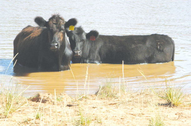
PHOTO 1: Avoid grazing wetland areas during peak culicoides activity periods. Photo provided by Beverly Moseley, USDA-NRCS Texas.
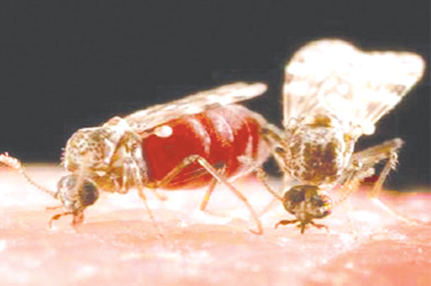
PHOTO 2: Biting midge feeding on a blood meal. Photo provided by Dr. Sonja Swiger, Texas A&M AgriLife Extension.
PHOTO 3: Culicoides larval habitats include mud, sand and debris at edges of ponds. Photo by Robert Fears.
Robert Fears is a freelance writer based in Georgetown, Texas. Email Robert Fears.


